
![on-carbon-fiber-types-and-their-carcinogenic-risks-[hackaday]](https://i0.wp.com/upmytech.com/wp-content/uploads/2024/08/202419-on-carbon-fiber-types-and-their-carcinogenic-risks-hackaday.jpg?resize=800%2C445&ssl=1)
On Carbon Fiber Types and Their Carcinogenic Risks [Hackaday]
Initially only seeing brief popular use as the filament in incandescent lighting, carbon fibers (CF) experienced a resurgence during the 20th century as part of composite materials that are lighter and stronger than materials like steel and aluminium, for use in aircraft, boats and countless more applications. This rising popularity has also meant that the wider population is now exposed to fragments of CF, both from using CF-based products as well as from mechanically processing CF materials during (hobby) projects.
It is this popularity that has also led to the addition of short CF sections to FDM 3D printing filaments, where they improve the mechanical properties of the printed parts. However, during subsequent mechanical actions such as sanding, grinding, and cutting, CF dust is created and some fraction of these particles are small enough to be respirable. Of these, another fraction will bypass the respiratory system’s dust clearing mechanisms, to end up deep inside the lungs. This raises the question of whether CF fragments can be carcinogenic, much like the once very popular and very infamous example of asbestos mineral fibers.
Making Carbon Fiber
The process of producing carbon fiber is fairly straightforward, involving a carbon-rich monomer, the precursor, that’s coaxed into becoming a polymer prior to having the non-carbon elements removed from the polymer. This then leaves the pure carbon, which depending on the precursor material will have certain physical and mechanical properties. The two most common precursor materials are polyacrylonitrile (PAN, (C3H3N)n) and pitch, the latter being a viscoelastic polymer that is either produced from petroleum or derived from plants. Both materials are used in many other applications as well.
In the case of CF production, the polymer is stretched and stabilized as part of pre-treatment, followed by carbonization and finally graphitization. In the case of PAN CF the precursor fiber is stabilized through oxidation while heated, followed by carbonization at much higher temperatures under an inert nitrogen atmosphere before undergoing a final third step called graphitization. This induces a graphite crystalline molecular structure arranged in a turbostratic (shifted planes) fashion and completing the CF which can then wound on to bobbins and readied for shipping.
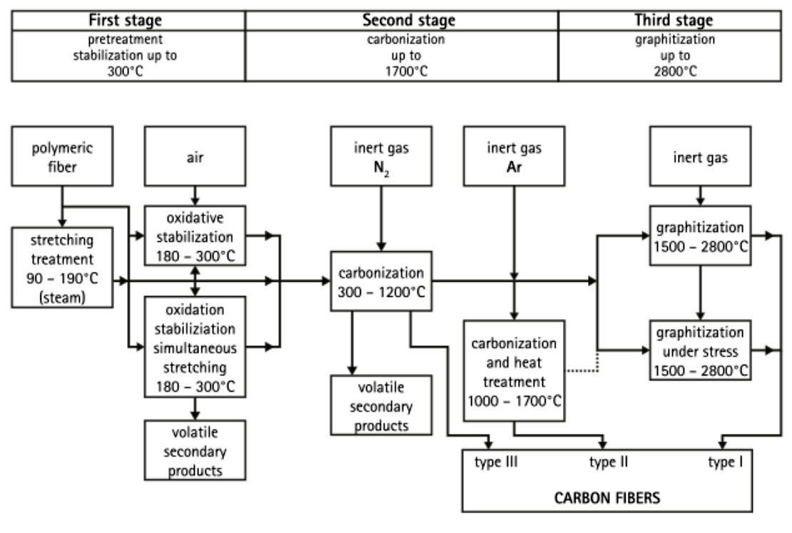
The turbostratic structure of PAN CF contrasts with the mesophase pitch CFs, which form strongly directional (sheet-like) graphitic structures. It is this difference in graphitic structure which ultimately determines the properties of PAN and pitch CF, such as a very high elastic modulus in the case of the latter and high tensile strength for the former. Currently the overwhelming majority of CF produced today is in the form of PAN CF, but pitch- and rayon-based CF are also used where their properties are superior to those of PAN CF.
CF is commonly used to create composites, in which case they are referred to as carbon fiber reinforced polymer (CFRP).
Creating Dust
That CF produces fragments when mechanically or thermally stressed should come as little surprise, with research on the health implications of CF fragments going back far into the 20th century. It is however only much more recently that we have been able to fully study the effects of CF fragments on lung tissue, not only based on empirical studies but also by looking at gene expression changes, DNA breakage, inflammation markers and so on.
Recently there was a bit of a furore surrounding the topic of CF fragments resulting from CF-infused FDM 3D printer filaments, with the question being raised of whether CF might be the new asbestos. In the linked article a 2017 paper by Jing Wang et al. as published in Journal of Nanobiotechnology was referenced. The researchers compared the way in which asbestos and carbon fiber fragments formed, as well as in the case of carbon nanotubes (CNTs).
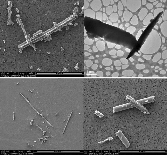
The SEM and TEM shown here on the right comes from a 2015 study by Schlagenhauf et al., in which PAN-based (Teijin IMS60) CFRP cable was exposed to extreme tension until failure. During this CFRP cable failure, many fragments were produced, some of which were deemed respirable according to the WHO criteria (minimum length of 5 µm and maximum diameter of 3 µm). This thus shows that PAN CF can in fact produce respirable fragments, but the pertinent question is whether these fragments are as harmful as asbestos fibers, or even pitch CF fragments.
In an article response on Twitter by Josef Prusa, the founder of Prusa 3D – a 3D printer and filament company – made the assertion that PAN-based filaments are in fact quite safe and that studies back this up, citing a 2019 study by Dominic Kehren et al. as published in Aerosol and Air Quality Research.
PSA
You might have seen the recent videos from @NathanBuilds or an article on @hackaday about the potential dangers of carbon fibers in filaments, comparing it to asbestos
Given that we offer several filaments containing carbon fibers, I thought many of you would be… pic.twitter.com/SjTTbqGe4N
— Josef Prusa (@josefprusa) August 4, 2024
This study by Kehren et al. looked at the amount of respirable fragments produced due to a limited number of mechanical actions. Their conclusion was that pitch CF fragments pose a significant health risk, including lung cancer and mesothelioma, while for PAN CF fragments more research was warranted. Of note here is also that no study was made of the actual effects of these biopersistent carbon fragments on lung tissue. Only assumptions of potential health implications were made based on WHO criteria and the observed fragments.
The advice by Josef Prusa to always wear a respirator while working with CF-based materials would thus seem to be rather prudent advice. Meanwhile we got much more recent studies which actually consider the physiological impact of exposure to PAN-based CF fragments.
In Vitro Lungs
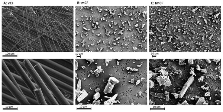
Whereas previous studies would generally use in vivo study models with all of the (ethical) complications which doing so entails, more recently in vitro models have become a viable alternative. In a 2023 study on PAN CF fragment effects on lung tissue by Alexandra Friesen et al. as published in International Journal of Molecular Sciences, a series of in vitro lung tissue models were used to directly study the effects of these fragments, including inflammation, DNA breakage and changes to gene expression.
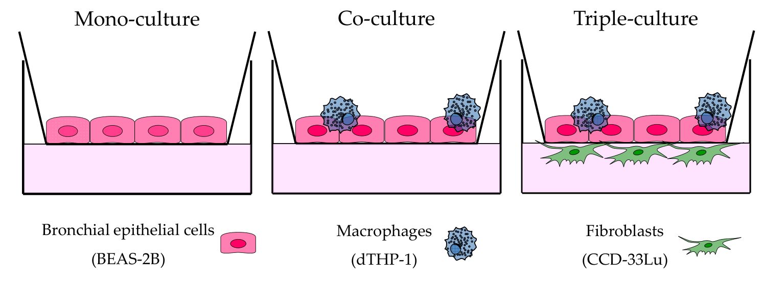 Schematic representation of the three cell culture models used for CF exposure experi-
ments. (Credit: Friesen et al., Int J Mol Sci, 2023)
These three different human cell culture models were exposed to both mechanically pre-treated (mCF) and thermo-mechanically pretreated (tmCF), using Teijin UMS40 filament yarn as the source of PAN CF. For both types of CF fragments the filament yarn was cut into 1 cm sections, which for mCF were immediately milled in a planetary ball mill. The tmCF pieces were first exposed to thermal stress in two furnaces (400°C for 4 hours, 800°C for ~30 minutes) in a nitrogen atmosphere before also being milled. The resulting fragments were then aerosolized and the cell culture models exposed.
After exposure, the cell cultures were post-incubated for 0, 3 or 23 hours to give an indication of how the exposure affects them over time. During exposure, the mCF showed a WHO fiber fraction of around 20%, whereas for tmCF this was around 9.4%. When analyzing the exposed cultures following post-incubation no significant cytotoxic effects were observed (based on LDH release).
Where things got more interesting was in the gene expression profiles, with the tmCF-exposed cultures showing the strongest pro-inflammation, cell-death (apoptosis) and DNA damage response. Interestingly, the triple-culture with fibroblasts present showed a less dramatic picture after 23 hours since mCF exposure, with inflammation transcription factors in particular significantly downregulated, though DNA damage response transcription was still upregulated.
Correspondingly, interleukin-8 (IL-8) release increased, though by an extraordinary level in the triple-culture. The exact reason for this and related observations remain to be investigated.
Genotoxicity was assessed after the increased DNA repair transcription, by analyzing DNA breaks. This provided the most interesting results, as it was found that while single-strand breaks were present in the mono- and co-culture samples, the triple-culture showed a significantly higher number of single-strand breaks. This indicates a genotoxic response upon exposure to PAN CF fragments, with the tmCF fragments showing the strongest response, possibly due to the production of more smaller fragments.
Although not as aggressively toxic as the multi-walled carbon nanotubes (MWCNTs) which the authors contrast with, the triple-culture results hint at secondary genotoxicity caused by the pro-inflammatory response.
Actions To Take
Although these in vitro cell culture models are not a perfect representation of human lungs, they do provide us with some of the first concrete evidence of how PAN CF can affect human lung tissue. With a clear inflammatory response that seems to get more significant and more harmful the more complete the cell culture becomes compared to a living lung’s insides, there is at the very least a solid basis to treat any CF dust as potentially carcinogenic.
Similar to asbestos fibers, it is not so much the material itself that causes toxicity and promotes carcinogenesis, but rather the body’s response to this foreign matter. Through sustained irritation, inflammation and subsequent DNA damage with possibly flawed repairs, outcomes such as silicosis, lung cancer and mesothelioma become possible.
The reasonable response here thus would be to treat CF no different from asbestos and similar sources of respirable, biopersistent fibers. This means not disturbing them if possible, being wary around damaged CFRP products and always wearing a respirator with asbestos-rated filters when sanding, cutting or otherwise processing CF in any form or shape, making sure to properly ventilate the room.
As with asbestos, it’s quite possible that being exposed will not cause permanent harm, but when the choice is between taking all possible safety precautions now, or finding out that you should have done so in one or two decades, it should hopefully be obvious what the reasonable choice is as we wait for more studies to be performed.